COMPARISON OF PRIMARY AND SECONPurpose: The aim of this study was to compare primary versus secondary forms of
multiple evanescent white dot syndrome (MEWDS) at T0 (baseline) and T1 (1–4 months
after the onset of symptoms).
Methods: A total of 101 eyes in 100 patients were included in a multicentric
retrospective study.
Results: Secondary MEWDS was defined as MEWDS associated with underlying
chorioretinal inflammatory pathologies, mainly multifocal choroiditis and punctuate inner
choroidopathy. Patients with secondary MEWDS were older (P = 0.011). The proportion of
women (P = 0.8), spherical equivalent (P = 0.3), and best-corrected visual acuity at T0 (P =
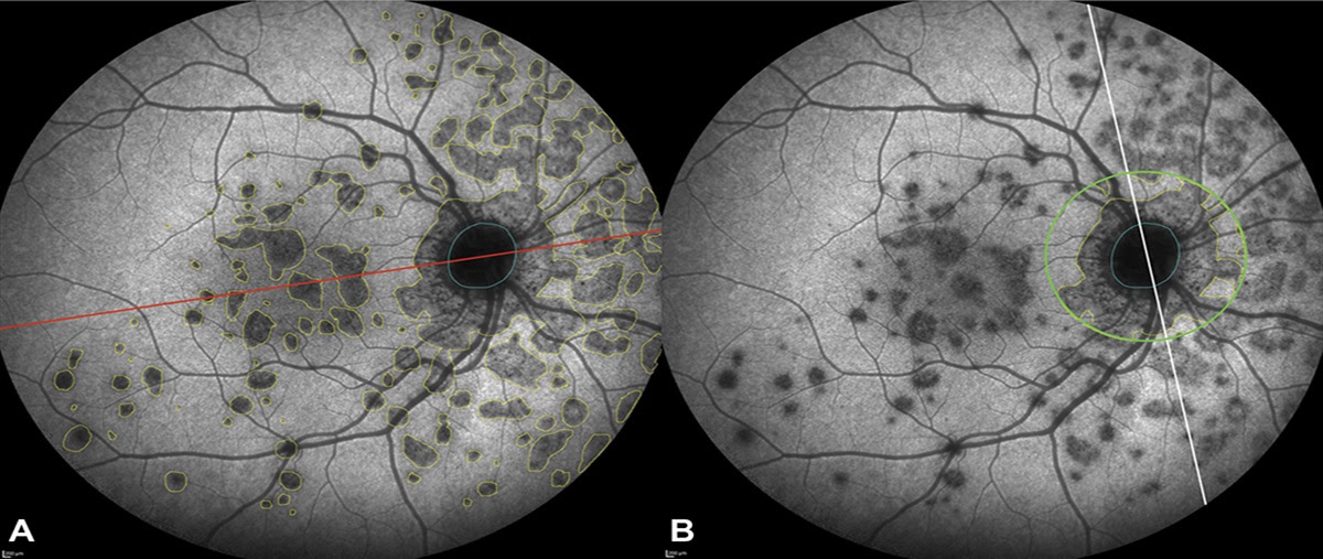
0.2) were not significantly different between the two groups. The area of MEWDS lesions on
late-phase indocyanine green angiography was significantly smaller in secondary MEWDS
(P = 0.001) and less symmetrical with respect to both horizontal (P = 0.003) and vertical (P =
0.004) axis. At T0, neither the clinical (P = 0.5) nor the multimodal imaging (P = 0.2)
inflammation scores were significantly different between the groups. At T1, the multimodal
imaging inflammation score was higher in secondary MEWDS (P = 0.021).
Conclusion: In secondary MEWDS, outer retinal lesions are less extensive and located
close to preexisting chorioretinal lesions. Mild signs of intraocular inflammation on
multimodal imaging are more frequent in secondary MEWDS during recovery. These
findings suggest that chorioretinal inflammation may trigger secondary MEWDS.
RETINA 42:2368–2378, 2022DARY FORMS OF MULTIPLE EVANESCENT WHITE DOT SYNDROME
Lire l’article : COMPARISON_OF_PRIMARY_AND_SECONDARY_FORMS_OF.17 (1)